News and Commentary Archive

Explore recent scientific discoveries and news as well as CLBB events, commentary, and press.

Mission

The Center for Law, Brain & Behavior puts the most accurate and actionable neuroscience in the hands of judges, lawyers, policymakers and journalists—people who shape the standards and practices of our legal system and affect its impact on people’s lives. We work to make the legal system more effective and more just for all those affected by the law.

WATCH — Boys to Men to Boys

clbb_boys2men2boys_poster_11x17_160411

Click poster to enlarge.

Approximately 2,000 youth sentenced to life without parole are now serving unconstitutional sentences in US prisons. What is the role for psychology and neuroscience in re-sentencing and parole after Miller and Montgomery?

Join two experts in forensic psychology, law and juvenile justice policy, for a discussion of the dilemmas posed after the Supreme Court’s recent decision to ban mandatory life without possibility of parole for juvenile homicides (Miller v. Alabama, 2012) and then this year to retroactively apply this decision to some 2,000 incarcerated individuals (Montgomery v. Louisiana, 2016).

The event will be held at 12:00 pm on Wednesday, April 13, in Wasserstein Hall, Milstein East C (2036) at Harvard Law School (1585 Massachusetts Avenue, Cambridge, MA).

This event is free and open to the public. Lunch will be served.

Continue reading »

WATCH — Poverty, Violence, and the Developing Mind

Click poster to RSVP.

Concentrated poverty is on the rise, and an increasing number of children are at risk for exposure to severe violence and dangerous living conditions. What are the implications of trauma exposure for healthy brain development?

During this panel event, Dr. Kerry Ressler (of McLean Hospital and Harvard Medical School) will discuss the risks poor, urban environments pose for post-traumatic stress disorder, while Dr. Charles A. Nelson (of Boston Children’s Hospital and Harvard Medical School) will discuss the effects of “toxic stress” on early childhood development. Carey Goldberg of WBUR will facilitate the conversation and host the Q&A session with the audience. 

This event will be held on Thursday, March 24, 2016, at the Brigham and Women’s Hospital, Bornstein Amphitheater, from 7:00-8:30 pm.

Make sure to RSVP before the event!

This event is free and open to the public. A brief reception will precede the event from 6:30-7:00 PM.

Continue reading »

Hyperactivity of Caudate, Parahippocampal, and Prefrontal Regions During Working Memory in Never-Medicated Persons at Clinical High-Risk for Psychosis

By Heidi W. Thermenos, Richard J. Juelich, Samantha R. DiChiara, Raquelle I. Mesholam-Gately, Kristen A. Woodberry, Joanne Wojcik, Nikos Makris, Matcheri S. Keshavan, Susan Whitfield-Gabrieli, Tsung-Ung W. Woo, Tracey L. Petryshen, Jill M. Goldstein, Martha E. Shenton, Robert W. McCarley, and Larry J. Seidman | Schizophrenia Research | March 7, 2016

Abstract:

Background

Deficits in working memory (WM) are a core feature of schizophrenia (SZ) and other psychotic disorders. We examined brain activity during WM in persons at clinical high risk (CHR) for psychosis.

Methods

Thirty-seven CHR and 34 healthy control participants underwent functional MRI (fMRI) on a 3.0 T scanner while performing an N-back WM task. The sample included a sub-sample of CHR participants who had no lifetime history of treatment with psychotropic medications (n = 11). Data were analyzed using SPM8 (2-back > 0-back contrast). Pearson correlations between brain activity, symptoms, and WM performance were examined.

Results

The total CHR group and medication-naive CHR sub-sample were comparable to controls in most demographic features and in N-back WM performance, but had significantly lower IQ. Relative to controls, medication-naïve CHR showed hyperactivity in the left parahippocampus (PHP) and the left caudate during performance of the N-back WM task. Relative to medication-exposed CHR, medication naïve CHR exhibited hyperactivity in the left caudate and the right dorsolateral prefrontal cortex (DLPFC). DLPFC activity was significantly negatively correlated with WM performance. PHP, caudate and DLPFC activity correlated strongly with symptoms, but results did not withstand FDR-correction for multiple comparisons. When all CHR participants were combined (regardless of medication status), only trend-level PHP hyperactivity was observed in CHR relative to controls.

Conclusions

Medication-naïve CHR exhibit hyperactivity in regions that subserve WM. These regions are implicated in studies of schizophrenia and risk for psychosis. Results emphasize the importance of medication status in the interpretation of task – induced brain activity.

Read the full article here.

Random Forest Segregation of Drug Responses May define Regions of Biological Significance

By Qasim Bukhari, David Borsook, Markus Rudin, and Lino Becerra | Frontiers in Computational Neuroscience | February 23, 2016

Abstract:

The ability to assess brain responses in unsupervised manner based on fMRI measure has remained a challenge. Here we have applied the Random Forest (RF) method to detect differences in the pharmacological MRI (phMRI) response in rats to treatment with an analgesic drug (buprenorphine) as compared to control (saline). Three groups of animals were studied: two groups treated with different doses of the opioid buprenorphine, low (LD) and high dose (HD), and one receiving saline. PhMRI responses were evaluated in 45 brain regions and RF analysis was applied to allocate rats to the individual treatment groups. RF analysis was able to identify drug effects based on differential phMRI responses in the hippocampus, amygdala, nucleus accumbens, superior colliculus and the lateral and posterior thalamus for drug vs. saline. These structures have high levels of mu opioid receptors. In addition these regions are involved in aversive signaling, which is inhibited by mu opioids. The results demonstrate that buprenorphine mediated phMRI responses comprise characteristic features that allow an unsupervised differentiation from placebo treated rats as well as the proper allocation to the respective drug dose group using the RF method, a method that has been successfully applied in clinical studies.

Read the full article here.

The Relations of Age and Pubertal Development with Cortisol and Daily Stress in Youth at Clinical Risk for Psychosis

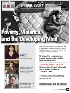
By Danielle M. Moskow, Jean Addington, Carrie E. Bearden, Kristin S. Cadenhead, Barbara A. Cornblatt, Robert Heinssen, Daniel H. Mathalon, Thomas H. McGlashan, Diana O. Perkins, Larry J. Seidman, Ming T. Tsuang, Tyrone D. Cannon, Scott W. Woods, and Elaine F. Walker | Schizophrenia Research | February 20, 2016

Abstract:

Background

Prodromal syndromes often begin in adolescence — a period of neurodevelopmental changes and heightened stress sensitivity. Research has shown elevated stress and cortisol in individuals at clinical high risk (CHR) for psychosis. This cross-sectional study examined relations of age and pubertal status with cortisol and self-reported stress in healthy controls (HCs) and CHR adolescents. It was hypothesized that the relations of age and pubertal stage with cortisol and stress would be more pronounced in CHR youth.

Methods

Participants were 93 HCs and 348 CHR adolescents from the North American Prodrome Longitudinal Study (NAPLS). At baseline, measures of stress (Daily Stress Inventory — DSI), Tanner stage (TS), and salivary cortisol were obtained.

Results

ANCOVA revealed increased DSI scores with age for both groups, and higher DSI scores in CHR adolescents than HCs, with a more pronounced difference for females. Contrary to prediction, with age controlled, HCs showed greater TS-related DSI increases. Analysis of cortisol showed no significant interactions, but a main effect of age and a trend toward higher cortisol in the CHR group. Correlations of cortisol with TS were higher in HC than CHR group.

Conclusions

Stress measures increased with age in HC and CHR adolescents, and DSI scores also increased with TS in HCs. The results do not support a more pronounced age or TS increase in stress measures in CHR adolescents, but instead suggest that stress indices tend to be elevated earlier in adolescence in the CHR group. Potential determinants of findings and future directions are discussed.

Read the full paper here.